Ear Attic Defect
Http Www Neurosurgeryresident Net Ear 20otology Ear38 20middle 20ear 20disorders Pdf

Mastoditis Middle Ear Head And Neck Sinusitis
Unit Four Middle Ear Disease Diagnosis

Hearing Disorders Chapter 20 The Cambridge Handbook Of Communication Disorders

Cholesteatoma Stanford Children S Health
Https Thieme Connect De Products Ebooks Pdf 10 1055 B 0039 171429 Pdf

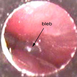
Overt attic cholesteatoma plus pars tensa collapse.
Ear attic defect.

Pin On My Posh Picks
Retracted Eardrum Symptoms Causes Diagnosis And Treatment

Zenith 7s 558 1940 Vintage Radio Antique Radio Old Radios

Getting Started In Endoscopic Ear Surgery Sciencedirect

Identify The Signs Hearing Problems Hearing Health Hearing Impairment

Bangs Thumb With Hammer Build A Closet Garage Door Opener Installation Attic Spaces

Cholesteatoma London Ear Clinic

Image Result For Scutum Erosion Facial Nerve Eustachian Tube Dysfunction Middle Ear

Marx Linemar Henry Eating Candy Windup Toy Vintage Toys Retro Toys Classic Toys

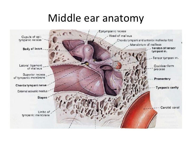
Anatomy Of Ear

Vintage Bunny Plush Rabbit Dakin Life Like Stuffed Animal Brown Tan Easter Dakin Korea 1986 In 2020 Bunny Plush Vintage Bunny Vintage Plush

Pdf Endoscopic Anatomy Of The Middle Ear

Time Out Area Study Area For The Easily Distracted Student With Some Of Those Defective I E Student Damaged Headphones As Ear Mufflers Small Space Office

The March Of Dimes Recommends Women Take These Six Simple Steps To Lower Their Babies Chances Of Having Birth Defects Birth Defects Lower Lake Baby

The Radiology Assistant Temporal Bone Anatomy 2 0 Cabeca E Pescoco Pescocinho

Vintage Louis Marx Roll Over Cat Wind Up Tin Toy W Box Ebay Igrushki
Https Www Bonalive Com Wp Content Uploads 2019 07 Mastoid Surgery Bonalive 9 100010 Pages Pdf

Development And Integration Of The Ear Sciencedirect
Https Encrypted Tbn0 Gstatic Com Images Q Tbn 3aand9gcsiselrebpbilelvtqodgauxo Kmsun7ny0i6wgwrf9mlvo Fnv Usqp Cau
Atticotmy

Vintage Mouse Figurine Vintage Mouse Knick Knack Mouse Mouse Eating Cheese Figurine Josef Originals 0 738 Wide Eyes Brow Vintage Collectibles Figurines

The Radiology Assistant Temporal Bone Anatomy 2 0 Cabeca E Pescoco Pescocinho

Vintage Anson Gold Tone Woven Rope Design Cufflinks Cufflinks Rope Design Woven
Https Www Healthplexus Net Files Content 2013 0306 0306cholesteatoma Pdf
Source : pinterest.com
